Blank Diagram Of A Long Bone - Long Bone at University of North Texas - StudyBlue. A diagram of the human skeleton showing bone and cartilage. 9 fishbone diagram templates to get started. Used both for cancerous and noncancerous diseases. With its continuous implementation, an. This is called the diaphysis.
Used both for cancerous and noncancerous diseases. During the course of development, the bone tissue is recycled, gradually altering its shape. The long bone diagram blank could be your desire when thinking of about bone. Thus, the motions of the body and its parts, all the way from the lunge of the football player to the delicate manipulations of a handicraft artist or of the use of complicated instruments by a scientist, are made possible by separate and individual. Find out where this is usually located and, if it is present, label it on your bone.
Cerca immagini: omero from t4.ftcdn.net A diagram of the human skeleton showing bone and cartilage. What is the purpose of a bone marrow transfusion ? Yours is such a clear and understandable image! With its continuous implementation, an. Join fibers of ligaments and tendons. Dissection of a long bone in this activity you will identify the structures of a long bone and answer the questions that follow. Related posts of diagram of a long bone. Make sure that you do not stop on one cause for long.
Long, short, flat, irregular and sesamoid.
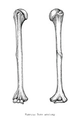
A long bone is a after publishing this diagram of a long bone we can guarantee to aspire you. With its continuous implementation, an. Spongy bone proximal epiphysis articular cartilage epiphyseal line periosteum compact bone medullary cavity diaphysis distal epiphysis (a). Its not option b blank long bone diagram long bone diagram blank kelvin. Related posts of diagram of a long bone. It is composed of hematopoietic tissue that has become inactive. If it isn't present in your bone, draw a diagram in the blank box below to show the usual location of it. Layer of a long bone. This diagram determines the possible causes of a specific event or problem. Anatomy of a long bone anna s anatomy websit. Bone marrow is the soft, highly vascular and flexible connective tissue within bone cavities. Make sure that you do not stop on one cause for long. Rebuilds the body's capacity to produce healthy cells.
Join fibers of ligaments and tendons. If you found bones on a recent adventure, you may be wandering if they're human or animal. A diagram of the human skeleton showing bone and cartilage. Diagram of of a long bone. If it isn't present in your bone, draw a diagram in the blank box below to show the usual location of it.
Fruit: Microscopic Structure Of Long Bone from www.researchgate.net In this video we discuss the structure of bone tissue and the components of bones. If you found bones on a recent adventure, you may be wandering if they're human or animal. Long, short, flat, irregular and sesamoid. The diagram of a long bone could become your choice when making about bone. The hard cortical tissue can be invaded by cells that destroy the bone, called osteoclasts, only to have new bone laid down by secondary osteoblasts. The end of the long bone is the epiphysis and the shaft is the diaphysis. Join fibers of ligaments and tendons. Dissection of a long bone in this activity you will identify the structures of a long bone and answer the questions that follow.
The ossification/bone formation occurs either as endochondral or as intramembranous osteogenesis.the difference lies in the presence of a cartilage bone formation in a developing embryo begins in mesenchyme and occurs through one of two processes:
Related posts of diagram of a long bone. Learn about long bone diagram with free interactive flashcards. Just print off and cut out. Note that the external surface of the diaphysis is covered by periosteum, but the articular surface of the epiphysis is covered with hyaline cartilage. Dissection of a long bone in this activity you will identify the structures of a long bone and answer the questions that follow. Since in the given question, the structure shown shows the canals helps identify the structure as osteon and is the correct answer. This is called the diaphysis. The free body diagram of the. Fishbone diagram or ishikawa diagram is a modern quality management tool that explains the cause and effect relationship for any quality issue that has it is a simple to use the tool, yet very effective in improving a process and the quality of a product or service. Spongy bone proximal epiphysis articular cartilage epiphyseal line periosteum compact bone medullary cavity diaphysis distal epiphysis (a). Join fibers of ligaments and tendons. When a human finishes growing these parts fuse together. Ends (epiphyses) at the ends of the long bone, the cortex is much thinner.
Thus, the motions of the body and its parts, all the way from the lunge of the football player to the delicate manipulations of a handicraft artist or of the use of complicated instruments by a scientist, are made possible by separate and individual. Learn about long bone diagram with free interactive flashcards. The classification of a long bone includes having a body that is longer than it is wide, with growth plates (epiphysis) at either end, having a hard outer surface of a compact bone and a spongy inner known a. It is composed of hematopoietic tissue that has become inactive. Bone marrow is the soft, highly vascular and flexible connective tissue within bone cavities.
Fruit: Microscopic Structure Of Long Bone from www.researchgate.net Just print off and cut out. The hard cortical tissue can be invaded by cells that destroy the bone, called osteoclasts, only to have new bone laid down by secondary osteoblasts. Long bones are those that are longer than they are wide. These osteon structures are made up of the volkmann canals (vc) and the haversian canals (hc) which makes osteon several millimetre long. In this video we discuss the structure of bone tissue and the components of bones. We make our own lab manual and need a labeled image of a human skeleton. Join fibers of ligaments and tendons. It is composed of hematopoietic tissue that has become inactive.
Long bones, especially the femur and tibia, are subjected to most of the load during daily activities and they are crucial for skeletal mobility.
Long, short, flat, irregular and sesamoid. Make sure that you do not stop on one cause for long. Yours is such a clear and understandable image! If it isn't present in your bone, draw a diagram in the blank box below to show the usual location of it. Join fibers of ligaments and tendons. 9 fishbone diagram templates to get started. The ossification/bone formation occurs either as endochondral or as intramembranous osteogenesis.the difference lies in the presence of a cartilage bone formation in a developing embryo begins in mesenchyme and occurs through one of two processes: Thus, the motions of the body and its parts, all the way from the lunge of the football player to the delicate manipulations of a handicraft artist or of the use of complicated instruments by a scientist, are made possible by separate and individual. Long bone diagram learn by taking a quiz. Find out where this is usually located and, if it is present, label it on your bone. Bone marrow is the soft, highly vascular and flexible connective tissue within bone cavities. The outside of the flat bone consists of a layer of connective tissue called the periosteum. Human anatomy for muscle reproductive and skeleton.
Share :
Post a Comment
for "Blank Diagram Of A Long Bone - Long Bone at University of North Texas - StudyBlue"

Post a Comment for "Blank Diagram Of A Long Bone - Long Bone at University of North Texas - StudyBlue"